
Let’s talk tiny. I mean really tiny. Like, get out your microscope small. I am talking about cells! You know them, you love them—those little bags of gobbledygook filled with smaller stuff still, with names like endoplasmic reticulum, ribosomes and Golgi bodies.
Cells are the building blocks of every living thing on the planet. So despite their size, they are kind of a big deal. So big that on a sunny Thursday afternoon I met up Marc Curtis, a cell biologist from Oregon State University, to talk about the small stuff.
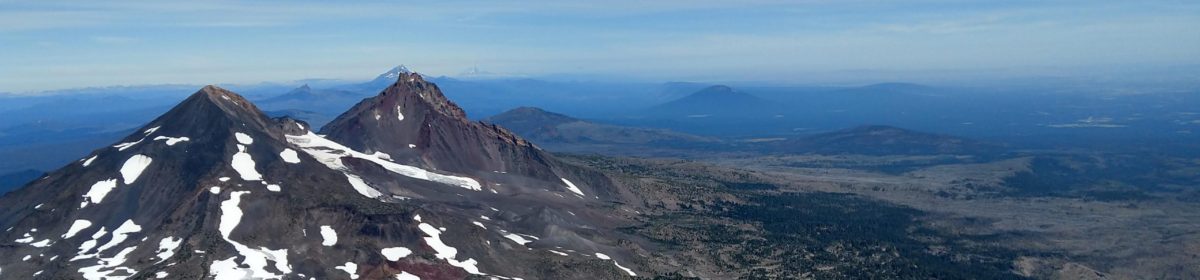
The Hike
- Trailhead: Forestry Club Cabin Trailhead at Peavy Arboretum, Corvallis, OR
- Distance: 4.8 miles
- Elevation Gain: approximately 900 feet
- Details: Plenty of parking at the trailhead. Restrooms available at the arboretum, but not at the trailhead. This hike is part of an extensive trail network throughout the McDonald-Dunn Forest, so there are many options to extend or shorten the hike.
Cell Level Thing
We immediately started hiking uphill from the parking lot along a forest road that leads to Oregon State Universities Forestry Club Cabin, before turning onto the tree-lined trail. As we trudged along several steep sections of the trail, Marc told me about his background.
Marc was fascinated by cells from a young age. In high school, his father, a Molecular Biologist at the Wistar Institute in Philadelphia, gave Marc a review article on cells and their role in cancer. Marc was hooked. “That just made sense to me, that cell level thing,” he said. “They grow, divide, they talk to each other, they take on different functions and they build the body.”
As an adult, Marc pursued his interest in cells by studying biochemistry at the University of New Hampshire and getting involved in undergraduate research involving cell signaling in the corpus luteum. Later, he moved onto Oregon State University to study cell death, as a graduate student, and eventually cell mutation as a postdoc.
Grow, Divide, Repeat
Marc has made a career out of thinking about cells, what they do, and how they do it. But cells are so small, invisible to the naked eye. Can we find a way to appreciate cells while on a hike?
A good start is to pay attention to plants. This is where Marc has focused most of his career.
Unlike humans, plants typically grow throughout their lives—packing on the inches at their tips, as long as conditions allow. Plant growth occurs in the roots and the shoots—where apical meristematic tissue composed of undifferentiated cells grow and divide. Meristematic tissue is also found in the buds and the nodes, the “joints” of a plant. Many different plant tissues and organs can arise from these growth regions, including leaves and flowers.
Marc pointed out the shoot apical meristem on one of the plants we saw along the trail. “It is in there,” he said, directing my eyes to the place where the leaf meets the stem. “That is where you have the undifferentiated cells—the fountain of youth—where new uncommitted cells come from.”
Marc explained that when these cells divide, they build up from the bottom layer and will eventually differentiate and become tissue, so the cells at the tips can maintain their undifferentiated status.

Time for a Change of Pace
However, this doesn’t mean these cells remain unaltered throughout the life of the plant. Because apical meristem divides so prolifically, mutations can accumulate in the meristematic tissue. Marc studied the process of mutation using meristematic tissue as a postdoc. Mutations are “mistakes” in DNA—a cell’s molecular instruction book—that arise during the replication process or from environmental factors, like UV light.
Marc was interested in how plant cells are able to bypass mutation so they can continue to grow and divide. Turns out that plant cells are pretty good at this. By maintaining a low level of fidelity during the replication process, cells in the apical meristem can continue their work of supplying the rest of the plant a lifetime supply of—well—cells! This also means there can be 100s of genetic variants in the meristem of one individual plant that can potentially give rise to unique growth forms. Though it seems like this is fairly rare.
In any event, growth is cellular! So when you see new leaves and flowers emerge in the spring, or look up to the tippy-top of a tree, or notice a new growth pattern in a familiar species—think tiny! Think— grow, divide, repeat! The leaves, flowers, and new growth each year are the result of the microscopic world of cellular division!

Beauty in Death
In autumn, cells take a turn for the morbid. As Marc and I made our way further up the trail, leaves crunching underfoot, I asked him to explain how cells were involved in the spectacular displays of fall foliage observed during this time of year.
The process is called autumn senescence—which essentially means a slow, seasonal death. Cells that make up the leaves of deciduous trees start to shut down in the fall in response to changes in daylight hours and temperature. In order to conserve resources, cells “break down chlorophyll and other components,” Marc explained, “leaving carotenoids and other pigments exposed.” Hence, the bright oranges, yellows, and purples. This organized way of dying, allows plants to hold onto difficult to obtain nutrients, like nitrogen, so that later in the spring they can begin to” grow, divide, repeat” once again.

Dead Tissue Eater
However, cells don’t always die in a blaze of colorful glory. Cell death may also be an adaptive defense. Earlier in the hike, Marc talked about his PhD work on a plant that when attacked by a toxic fungus would respond by activating cell death. This might seem like a bad idea at first glance, but by killing off the tissue where the fungi attacked, the plant was able to stave off further damage and prevent the fungus from eating it. You see, this particular fungi was a biotroph, meaning it only consumes living tissue. Dead tissue was entirely unappealing.
Unfortunately, Marc’s tale has a sad ending. Another fungus came along—a necrotroph, or dead tissue eater—that was able to mimic the biotrophic fungi’s toxin, triggering cell death in the plant. Only in this case, the dead tissue was very appetizing to the fungi. It is a dog eat dog cellular world out there!
Gall-y
Eventually, Marc and I made it to the top of the Powder House Trail, where the vegetation changed from the Douglas-fir and Big Leaf Maple trees we had spent most of our hike walking through, to a hilltop prairie of grasses and Oaks. Walking amidst the oak trees reminded me of the dozens of oak galls my kiddos and I had spotted amongst the fall foliage on another recent hike in the area. I asked Marc about these odd growths.
Though he didn’t know a lot of details, Marc did recall that the growth was the result of a parasitic “gall wasp.” The gall wasp will lay its eggs on an oak leaf or twig, usually in the spring. Then, when these eggs hatch into larvae, they release biochemicals that “brainwash” the cells of the leaf or twig into forming a gall. The gall—a growth filled with nutritive cells—envelops the larvae as it forms, protecting and feeding it while it pupates.
Upon further research, I found that gall shape and structure are unique to each parasite, even between individuals of the same species. And that the growth pattern of galls are so different from normal leaf or branch tissue growth, that “they have been described as new plant organs” with a unique chemical signature. Though it seems there is still more research needed to understand exactly how the larvae control cell growth and division, galls are an impressive example of what I like to call “zombie biology.”
Green Islands
In discussing galls, Marc was reminded of another parasitic relationship that can be observed in the fall—green islands. Green islands are spots on a leaf that will remain green even as the rest of the leaf begins to undergo senescence. Marc explained—the green spots are the result of a fungal infection. “The fungus produces plant hormones called cytokinin which delays senescence.” This keeps the chlorophyll—the green stuff involved in photosynthesis—from breaking down. Hence the green. This also means the plant’s food factory stays open for business, providing the fungus a continuous supply of sugar. Needless to say, this is a pretty sweet deal for the fungi.

Wax on, Wax off
As Marc and I meandered along the trail looking for green islands (later we found a few examples), Marc pointed toward a gnarled and twisted tree trunk with peeling red bark straight ahead—a Madrone!
A personal favorite of Marc’s I walked up to get a closer look. I felt one of the thick, smooth leaves. “What is going on here?” I asked. “Why do Madrone’s have waxy leaves?” It must be something cellular I thought.
I was right! “Waxes are secreted by the epidermal cells through the endomembrane system,” Marc replied. The endomembrane system is the machinery in a cell that packages and transports certain molecules, like wax, into the extracellular world. Marc explained, The Golgi probably synthesizes the wax. It is then gathered in vesicles. These vesicles transport the wax to the outer cellular membrane. Here they fuse with the membrane and dump the wax out into the cell wall. And voila—waxy leaves!
Human bone-building also uses the endomembrane system, Marc shared. Though in this case, the bone cells are spewing out collagen protein—the number one ingredient for bones. Think about it! Our skeletons are made from cells throwing up all day. A fun fact Marc likes to share with his students.

Evolution
Eventually, Marc and I made our descent back towards our cars. On our way down the trail, we chatted about topics ranging from teaching on zoom to plant podcasts.
Marc was also on the lookout for a liverwort he had been able to identify recently and wanted to see if he could find it again. In addition to several botany courses, Marc also teaches an evolution course at OSU. So with ancient plants on the brain, biological evolution naturally came up as we hiked along.
Innovation
Like most things, if you haven’t caught on yet, evolution is very much a cellular process. Mutations that occur in reproductive cells, gametes, provide new genetic variation to a population. Meiosis, the development of reproductive cells, does the same by mixing and matching genes so each gamete is unique.
Of course, there is much more to evolution by natural selection than genetic variation. When passing by a Douglas-fir tree, Marc shared with me his thoughts on the subject. “Bark,” he said, “has a high surface area with all the cracks and creases.” When bark like this evolved it provided opportunities for many other species to evolve on it. “Innovation opens the door for more innovation,” Marc explained. He used the analogy of the internet boom. Once the internet got started it provided opportunities for online retail, social media, etc. One new idea brought about many more ideas. Life is similar—a new biological innovation can open up ecological space for new species to emerge.

Listen to a Liverwort
Toward the end of our hike, Marc finally found his liverwort. He pointed out how the “leaf” structure and arrangement was different from that of a moss or any other plant. A difference that Marc had only recently developed an eye for, and I had never considered. In fact, I was pretty sure I have been mistaking liverwort for some other group, like a lichen or moss, my entire life.

Back at home, I kept thinking about Marc’s liverwort and his thoughts on innovation. And maybe because the world seems so divided, or perhaps it is a personal crisis of faith in mankind, but I can’t help but feel like there is some sort of deeper message here.
Throughout our hike, the concept of curiosity being essential to scientific work kept coming up. But I think it goes beyond science. We all need to be curious. Open our eyes to the liverworts of the world—not lump them together with lichen or a moss—assuming they are all the same. We need an evolution of the mind! And just like the bark of a tree, innovative ideas will open the mind to more innovative ideas.
Cells are amazing in their simple mantra—grow, divide, repeat. But it is mutation, change, or innovation—that keeps things moving forward— evolving.
So, let “new variations” or ideas sink in and become part of your mental framework. If nothing else, you may finally learn to identify a liverwort.
Marc Curtis is an instructor at Oregon State University in the Department of Botany and Plant Pathology. Marc has a Bachelor’s degree from the University of New Hampshire in Biochemistry and PhD from Oregon State University where he studied mutation in plant cells.
Very nice and honestly inspiring article when it came down to it. Next time I am on a hike or just a little walk around the neighborhood I am for sure going to pay more attention to the amazing thing mentioned here.